
Rheinberg Illumination
Click here for an overview of Rheinberg Illumination.
Click here to learn about the optical theory of Rheinberg Illumination.
Click here to learn about commercial Rheinberg solutions.
Click here to learns some practical tips for Rheinberg Illumination.

What Is Rheinberg Illumination?
Rheinberg Illumination, which was invented in 1986 by British microscopist Julius Rheinberg, is an optical staining technique derived from darkfield microscopy, wherein the simple patch stop used in low-power darkfield is replaced by a concentric, two-colored transparent gelatin or glass filter to provide contrasting colors to the specimen and background. Like darkfield, Rheinberg Illumination is used to provide enhanced contrast to live/unstained specimens that are difficult to see under normal brightfield conditions.
Theory of Rheinberg Illumination
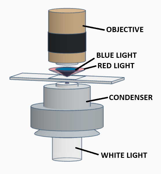
Rheinberg Illumination uses a two-colored concentric filter, consisting of a central disk of one color, surrounded by an outer (annular) ring of a second, contrasting color. This filter modifies the light cone produced by the substage condenser, imparting the color of the central disk to the light in the central portion of the cone, and the color of the annular ring to the light in the outer portion of the cone. The sizing of the central disk and the annular ring are chosen such that some light in the central portion of the cone will be directly collected by the numerical aperture of the objective, while the light in the outer portion will fall outside the collection angle of the objective and not contribute to the image (when there is no specimen on the stage). This results in a visual field brightly illuminated in the color of the central disk of the Rheinberg filter.
Let’s now consider what happens to the light in the outer portion of the cone when a specimen is placed on the stage. This light strikes the specimen and is scattered by diffraction, such that some of this scattered light will now fall into the collection angle of the objective and be collected. Since the image seen through the eyepieces is the result of wave interference at the intermediate image plane of the collected background light (of one color) and diffracted light (of a contrasting color), the specimen will appear in the color of the annular ring, against a background colored by the central disk.
Commercial Rheinberg
In the 1930s, The Carl Zeiss company manufactured an aplanatic condenser with three independent diaphragms, known as the Mikropolychromar, which was designed to produce Rheinberg images. The outermost diaphragm of the Mikropolychromar controlled the field diameter, while the two smaller diaphragms controlled the light passing through the central disk. The Mikropolychromar was supplied with a set of transparent annular rings and central disks of various colors. This highly collectable condenser is very rare, and is almost impossible to find on the used market.
A Few Practical Considerations
Be sure to always run the condenser diaphragm wide open when using Rheinberg Illumination.
- You may find it helpful to stack two or three pieces of the same color to make the central filter or annular ring, to get the desired depth of color from the filter.
- If you wish to decrease the intensity of the background, you can reduce the diameter of the central disk, but be careful. The size of the hole in the annular ring of the filter must be sized to keep the outer portion of the light cone outside the acceptance angle of the objective, regardless of the diameter of the central disk. This means if you wish to make the central disk smaller then the minimum diameter of the hole in the annular ring, you will need to place an opaque ring between the central disk and the annular ring.
- In general, the central filter should be much darker than the annular ring filter, to provide sharp contrast between the image and the background.