
Darkfield Microscopy
Click here to learn the optical theory of Darkfield Illumination.
Click here to learn about darkfield condensers.
Click here to learn about DIY darkfield.
Click here to learn how to add Darkfield Illumination to an Olympus BH2 microscope.
Click here to see a demonstration of the darkfield illumination cone.
Darkfield Illumination
Darkfield Illumination is an optical staining technique used to provide enhanced contrast to live/unstained specimens, such as diatoms or other single celled organisms, that are difficult to see under normal brightfield conditions. In simple terms, darkfield illumination illuminates the specimen in such a way that direct light is not collected by the objective lens, and only diffracted light which has been scattered by the specimen is collected to contribute to the final image. This exclusion of zero-order un-diffracted light and the collection of diffracted light produces a visual field wherein the specimen appears brightly illuminated against a dark background.
For most applications in professional microscopy, darkfield has largely been replaced by more modern imaging modes such as phase contrast and differential interference contrast, but darkfield is still useful for some specific applications. For example, darkfield is used in the diagnosis of certain diseases caused by spirochetes, which become quite visible in peripheral blood smears under darkfield illumination. Darkfield is also frequently used in the visualization of surface imperfections when using incident-light (reflected) microscopy.
Which Type of Darkfield Condenser Should I Use?
There are two types of dedicated darkfield condensers available. The first type, known as a dry darkfield condenser, is suitable for lower power objectives and does not use immersion oil. The second type, known as a wet darkfield condenser, is suitable for higher power objectives and requires the use of immersion oil between the top element of the condenser and the bottom of the specimen slide.
A dry darkfield condenser can typically be used with 10X through 20X objectives (maybe even 40x) and is much simpler and more convenient to use than a wet darkfield condenser. A wet darkfield condenser can typically be used with 20X through 100X objectives, but the requirement for immersion oil makes using them messy and much less convenient than a dry darkfield condenser.
Note that even if you have a wet darkfield condenser, you will not be able to obtain darkfield with a 100X oil-immersion objective unless your objective has an internal iris diaphragm to allow you to reduce the numerical aperture of the objective to below that of the illuminating cone. A 100x dry objective will not contain an iris diaphragm, as this is not needed for darkfield with a wet darkfield condenser.
DIY Darkfield
The easiest way to implement simple darkfield in a typical upright compound scope is to place an opaque disk, known as a darkfield patch stop, in the optical pathway (near the iris diaphragm of the condenser) on your scope. The presence of this patch stop blocks the central portion of light from passing through the condenser iris and thereby causes the condenser to produce a hollow cone of light for the specimen. See the image below for a comparison of normal brightfield Abbe condenser (shown on left) and the same condenser with a simple patch stop for darkfield (shown on right).
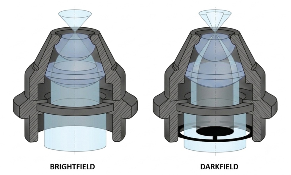
As can be seen in the image above, the hollow geometry of the darkfield lighting cone ensures that no direct light will pass through the slide and be collected by the objective lens. The oblique light that does strike the specimen would normally fall outside the collection angle of the objective lens, but some of this will be scattered by diffraction as it passes through the specimen. That which is sufficiently scattered to fall within the acceptance angle of the objective lens will be collected to contribute to the final image. The resulting image, which is produced by wave interference of the various orders of diffracted light collected by the objective lens, will reveal the classical darkfield image we all know and love (i.e., a brightly illuminated specimen against a dark background).
For best darkfield imaging, the patch stop should be placed as near to the iris diaphragm within the condenser as possible, and it must be centered relative to the diaphragm. The diameter of the patch stop must be sufficient to block all direct light that would otherwise be collected by the objective lens, and the iris diaphragm should be set to the fully open position.
If the diameter of the patch stop is too small, it will not exclude all of the un-scattered light and the specimen will not appear against an acceptably dark field. If the stop is too large, it will be hard to collect sufficient scattered light for an acceptably bright image. The ideal stop size would be such that the dark central portion of the lighting cone is just large enough that the objective does not see direct light, but not any larger. This of course means that the ideal stop size is a function of the numerical aperture of the objective. Each objective should, at least in theory, have a specific stop size for optimal darkfield performance. In practice, a single stop is typically used that is sized for the objective lens with the highest numerical aperture for which it will be used.
A simple darkfield stop is typically suitable for objective lenses ranging from 10X to 20X, and sometimes even 40X, but is never suitable for 100X. For proper darkfield at 40X, a dedicated darkfield condenser is often necessary, and for darkfield at 100X, a dedicated darkfield condenser is a necessity (as is an objective lens with an internal iris diaphragm to reduce the NA).
One should be careful when interpreting darkfield images. Many features seen in brightfield will be invisible in darkfield, especially those represented by low spatial frequencies (caused by the lack of direct light and the lowest diffraction orders in the collected light), making the image appear somewhat “high-passed”.
One well-known limitation of darkfield microscopy is the low light levels seen in the final image. Since most of the illumination available in conventional brightfield is obscured by the darkfield patch stop, the specimen must be very strongly illuminated in order to collect sufficient scattered light for an acceptable darkfield image, which can damage the specimen in some cases.
Darkfield images are not simply negative versions of the brightfield image. Different effects are visible in each type of illumination. In brightfield, some features can be seen where a shadow is cast on the surface by the incident light or where a part of the surface is less reflective. Raised features too smooth to cast shadows will not generally appear in brightfield images, but will be visible in the dark-field images.